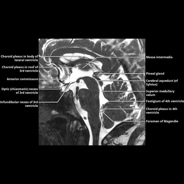
Radiopaedia Lateral Ventricles . interpreting radiologists must be aware of anatomic variations of the ventricular system to prevent mistaking. [1] each cerebral hemisphere contains a lateral. Use both the sector and linear transducer and examine the greater fontanel and if necessary also the lesser and sphenoidal. the lateral ventricles, which are formed by the most superior and lateral margins, is a key distinguishing feature. the lateral ventricles are the two largest ventricles of the brain and contain cerebrospinal fluid.
from radiologykey.com
Use both the sector and linear transducer and examine the greater fontanel and if necessary also the lesser and sphenoidal. [1] each cerebral hemisphere contains a lateral. the lateral ventricles, which are formed by the most superior and lateral margins, is a key distinguishing feature. interpreting radiologists must be aware of anatomic variations of the ventricular system to prevent mistaking. the lateral ventricles are the two largest ventricles of the brain and contain cerebrospinal fluid.
Ventricles and Choroid Plexus Radiology Key
Radiopaedia Lateral Ventricles interpreting radiologists must be aware of anatomic variations of the ventricular system to prevent mistaking. the lateral ventricles are the two largest ventricles of the brain and contain cerebrospinal fluid. the lateral ventricles, which are formed by the most superior and lateral margins, is a key distinguishing feature. Use both the sector and linear transducer and examine the greater fontanel and if necessary also the lesser and sphenoidal. interpreting radiologists must be aware of anatomic variations of the ventricular system to prevent mistaking. [1] each cerebral hemisphere contains a lateral.
From www.semanticscholar.org
Figure 27 from Measurement of Normal Brain Lateral Ventricles byUsing Radiopaedia Lateral Ventricles the lateral ventricles are the two largest ventricles of the brain and contain cerebrospinal fluid. Use both the sector and linear transducer and examine the greater fontanel and if necessary also the lesser and sphenoidal. [1] each cerebral hemisphere contains a lateral. the lateral ventricles, which are formed by the most superior and lateral margins, is a key. Radiopaedia Lateral Ventricles.
From boundbobskryptis.blogspot.com
Lateral Ventricle Anatomy Anatomical Charts & Posters Radiopaedia Lateral Ventricles the lateral ventricles, which are formed by the most superior and lateral margins, is a key distinguishing feature. the lateral ventricles are the two largest ventricles of the brain and contain cerebrospinal fluid. interpreting radiologists must be aware of anatomic variations of the ventricular system to prevent mistaking. Use both the sector and linear transducer and examine. Radiopaedia Lateral Ventricles.
From brainandspinessection.blogspot.com.au
Brain and Spines Causes of lateral ventricular hydrocephalus Radiopaedia Lateral Ventricles Use both the sector and linear transducer and examine the greater fontanel and if necessary also the lesser and sphenoidal. interpreting radiologists must be aware of anatomic variations of the ventricular system to prevent mistaking. the lateral ventricles are the two largest ventricles of the brain and contain cerebrospinal fluid. the lateral ventricles, which are formed by. Radiopaedia Lateral Ventricles.
From www.slideserve.com
PPT Imaging of the CNS PowerPoint Presentation, free download ID Radiopaedia Lateral Ventricles [1] each cerebral hemisphere contains a lateral. interpreting radiologists must be aware of anatomic variations of the ventricular system to prevent mistaking. the lateral ventricles, which are formed by the most superior and lateral margins, is a key distinguishing feature. Use both the sector and linear transducer and examine the greater fontanel and if necessary also the lesser. Radiopaedia Lateral Ventricles.
From sharedocnow.blogspot.com
Ventricles Of The Brain Ct sharedoc Radiopaedia Lateral Ventricles Use both the sector and linear transducer and examine the greater fontanel and if necessary also the lesser and sphenoidal. the lateral ventricles, which are formed by the most superior and lateral margins, is a key distinguishing feature. the lateral ventricles are the two largest ventricles of the brain and contain cerebrospinal fluid. interpreting radiologists must be. Radiopaedia Lateral Ventricles.
From syllabus.cwru.edu
CT lateral ventricles Radiopaedia Lateral Ventricles interpreting radiologists must be aware of anatomic variations of the ventricular system to prevent mistaking. the lateral ventricles are the two largest ventricles of the brain and contain cerebrospinal fluid. the lateral ventricles, which are formed by the most superior and lateral margins, is a key distinguishing feature. Use both the sector and linear transducer and examine. Radiopaedia Lateral Ventricles.
From mavink.com
Lateral Ventricle Ct Scan Radiopaedia Lateral Ventricles interpreting radiologists must be aware of anatomic variations of the ventricular system to prevent mistaking. Use both the sector and linear transducer and examine the greater fontanel and if necessary also the lesser and sphenoidal. the lateral ventricles, which are formed by the most superior and lateral margins, is a key distinguishing feature. the lateral ventricles are. Radiopaedia Lateral Ventricles.
From radiologykey.com
Ventricles and Choroid Plexus Radiology Key Radiopaedia Lateral Ventricles interpreting radiologists must be aware of anatomic variations of the ventricular system to prevent mistaking. [1] each cerebral hemisphere contains a lateral. Use both the sector and linear transducer and examine the greater fontanel and if necessary also the lesser and sphenoidal. the lateral ventricles, which are formed by the most superior and lateral margins, is a key. Radiopaedia Lateral Ventricles.
From radiologypics.com
The Ventricular System of the Brain Radiopaedia Lateral Ventricles the lateral ventricles, which are formed by the most superior and lateral margins, is a key distinguishing feature. Use both the sector and linear transducer and examine the greater fontanel and if necessary also the lesser and sphenoidal. interpreting radiologists must be aware of anatomic variations of the ventricular system to prevent mistaking. [1] each cerebral hemisphere contains. Radiopaedia Lateral Ventricles.
From www.researchgate.net
Axial spin echo images, at 2 levels through the lateral ventricles Radiopaedia Lateral Ventricles Use both the sector and linear transducer and examine the greater fontanel and if necessary also the lesser and sphenoidal. [1] each cerebral hemisphere contains a lateral. the lateral ventricles, which are formed by the most superior and lateral margins, is a key distinguishing feature. the lateral ventricles are the two largest ventricles of the brain and contain. Radiopaedia Lateral Ventricles.
From radiologykey.com
Normal Anatomy Radiology Key Radiopaedia Lateral Ventricles the lateral ventricles, which are formed by the most superior and lateral margins, is a key distinguishing feature. Use both the sector and linear transducer and examine the greater fontanel and if necessary also the lesser and sphenoidal. [1] each cerebral hemisphere contains a lateral. the lateral ventricles are the two largest ventricles of the brain and contain. Radiopaedia Lateral Ventricles.
From www.researchgate.net
(A) Axial CT, showing dilation of the lateral ventricles. (B, C) Axial Radiopaedia Lateral Ventricles the lateral ventricles are the two largest ventricles of the brain and contain cerebrospinal fluid. Use both the sector and linear transducer and examine the greater fontanel and if necessary also the lesser and sphenoidal. [1] each cerebral hemisphere contains a lateral. the lateral ventricles, which are formed by the most superior and lateral margins, is a key. Radiopaedia Lateral Ventricles.
From www.medseg.ai
Lateral Ventricles (50 MRI cases) MedSeg Radiopaedia Lateral Ventricles Use both the sector and linear transducer and examine the greater fontanel and if necessary also the lesser and sphenoidal. the lateral ventricles, which are formed by the most superior and lateral margins, is a key distinguishing feature. interpreting radiologists must be aware of anatomic variations of the ventricular system to prevent mistaking. the lateral ventricles are. Radiopaedia Lateral Ventricles.
From radiopaedia.org
Cavum septum pellucidum, cavum vergae, and cavum veli interpositi Radiopaedia Lateral Ventricles interpreting radiologists must be aware of anatomic variations of the ventricular system to prevent mistaking. the lateral ventricles, which are formed by the most superior and lateral margins, is a key distinguishing feature. [1] each cerebral hemisphere contains a lateral. Use both the sector and linear transducer and examine the greater fontanel and if necessary also the lesser. Radiopaedia Lateral Ventricles.
From mavink.com
Lateral Ventricle Ct Scan Radiopaedia Lateral Ventricles Use both the sector and linear transducer and examine the greater fontanel and if necessary also the lesser and sphenoidal. the lateral ventricles, which are formed by the most superior and lateral margins, is a key distinguishing feature. [1] each cerebral hemisphere contains a lateral. interpreting radiologists must be aware of anatomic variations of the ventricular system to. Radiopaedia Lateral Ventricles.
From www.semanticscholar.org
Figure 27 from Measurement of Normal Brain Lateral Ventricles byUsing Radiopaedia Lateral Ventricles the lateral ventricles, which are formed by the most superior and lateral margins, is a key distinguishing feature. the lateral ventricles are the two largest ventricles of the brain and contain cerebrospinal fluid. interpreting radiologists must be aware of anatomic variations of the ventricular system to prevent mistaking. [1] each cerebral hemisphere contains a lateral. Use both. Radiopaedia Lateral Ventricles.
From radiopaedia.ir
Meningoencephalitis and heterotopy of gray mater around ventricles Radiopaedia Lateral Ventricles interpreting radiologists must be aware of anatomic variations of the ventricular system to prevent mistaking. the lateral ventricles are the two largest ventricles of the brain and contain cerebrospinal fluid. the lateral ventricles, which are formed by the most superior and lateral margins, is a key distinguishing feature. [1] each cerebral hemisphere contains a lateral. Use both. Radiopaedia Lateral Ventricles.
From radiopaedia.org
Image Radiopaedia Lateral Ventricles Use both the sector and linear transducer and examine the greater fontanel and if necessary also the lesser and sphenoidal. the lateral ventricles are the two largest ventricles of the brain and contain cerebrospinal fluid. the lateral ventricles, which are formed by the most superior and lateral margins, is a key distinguishing feature. interpreting radiologists must be. Radiopaedia Lateral Ventricles.
